https://creativecommons.org/publicdomain/zero/1.0/https://www.rawpixel.com/image/9976792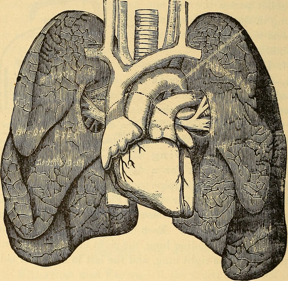
Identifier: anatomyphysiolog00mayc (find matches)Title: Anatomy, physiology and hygieneYear: 1890 (1890s)Authors: May, Charles Henry, 1861-1943Subjects: Human anatomy Physiology Hygiene, Popular. (from old catalog)Publisher: New York, W. Wood and companyContributing Library: The Library of CongressDigitizing Sponsor: The Library of CongressView Book Page: Book ViewerAbout This Book: Catalog EntryView All Images: All Images From BookClick here to view book online to see this illustration in context in a browseable online version of this book.Text Appearing Before Image:The Air-passage and the Food-passage. The heavy line indicates the courseof the food through mouth and gullet; thedotted line shows the course of air throughnostril into pharynx, and then into the lar-ynx and trachea, which are placed in front ofthe gullet. THE LUNGS. 279. The lungs are the organs with which we breathe. Theentire lung may be divided into two halves (Figs. 68 and 69),a right lung and a left lung. Between these two the heart isplaced (Fig. 68). The lungs and the heart fill up the entirespace in the chest. 280. Shape of the Lungs.âEach lung is shaped some-what like a cone, with the apex above and the base below (Fig. 136 ANATOMY, PHYSIOLOGY, AND HYGIENE. 69). The lungs are very light and contain a great deal of air ;they float when placed on water. Even after squeezing out allthe air we can, there will still be a considerable quantity re-maining in the lung. 281. Structure of the Lungs.âIf we cut into the lungs,we find they are formed of a large number of tubes andText Appearing After Image:Fig. 68.âThe Heart and Lungs. On each side the lungs are seen ; in the centre is theheart; above are the windpipe, and the laige blood-vessels passing to and from the heart. spaces containing air. After entering the lungs each bron-chus divides again and again (Fig. 69), each branch, knownas a bronchial tube, becoming smaller, until finally thebranches of each bronchial tube have become so small thatthey can no longer be seen without the microscope (Fig.70, a). THE ORGANS OF VOICE AND BREATHING. 137Note About ImagesPlease note that these images are extracted from scanned page images that may have been digitally enhanced for readability - coloration and appearance of these illustrations may not perfectly resemble the original work.
Original public domain image from Wikimedia Commons
Public DomainFree CC0 image for Personal and Business use