https://creativecommons.org/publicdomain/zero/1.0/https://www.rawpixel.com/image/9976740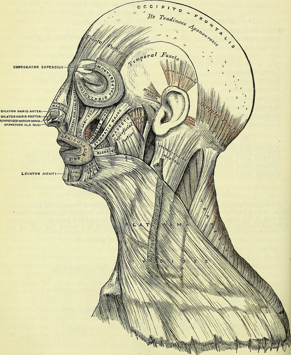
Identifier: b20386424 (find matches)Title: Anatomy, descriptive and surgical (electronic resource)Year: 1860 (1860s)Authors: Gray, Henry, 1825-1861 Carter, H. V., ill Westmacott, John Guise, Dr, illSubjects: AnatomyPublisher: London : J.W. ParkerContributing Library: Wellcome LibraryDigitizing Sponsor: Wellcome LibraryView Book Page: Book ViewerAbout This Book: Catalog EntryView All Images: All Images From BookClick here to view book online to see this illustration in context in a browseable online version of this book.Text Appearing Before Image:2. 3. of AUniCULAR RECIOIM J^.S.G.qf FACE^,8. of NECK head, from the anterior to the posterior extremity of the preceding. Raise the skin infront from the subjacent muscle from below upwards; this must be done with extremecare, on account of their intimate union. The tendon of the muscle is best avoided byremovmg the integument from the outer surface of the vessels and nerves which hebetween the two. 208 MUSCLES AND FASCIA. The superficial fascia in the epicranial region is a firm, dense layer, intimatelyadherent to the integument, and to the Occipito-frontalis and its tendinous aponeu-rosis; it is continuous, behind, with the superficial fascia at the back part of theneck; and, laterally, is continued over the temporal aponeurosis: it containsText Appearing After Image:148.—Muscles of the Head, Face, and Neck. between its layers the small muscles of the auricle, and the superficial temporalvessels and nerves. The Occipito-frontalis (fig. 148) is a broad musculo-fibrous layer, which coversthe whole of one side of the vertex of the skull, from the occiput to the eyebrow.It consists of two muscular slips, separated by an intervening tendinous aponeurosis.The occipital portion^ thin, quadrilateral in form, and about an inch and a half in AURICULAR REGION. 209 length, arises from the outer two-thirds of the superior curved line of the occipitalbone, and from the mastoid portion of the temporal. Its fibres of origin aretendinous, but they soon become muscular, and ascend in a parallel direction toterminate in the tendinous aponeurosis. The frontal portion is thin, of aquadrilateral form, and intimately adherent to the skin. It is broader, its fibresare longer, and their structure paler than the occipital portion. Its internal fibresare continuous with tNote About ImagesPlease note that these images are extracted from scanned page images that may have been digitally enhanced for readability - coloration and appearance of these illustrations may not perfectly resemble the original work.
Original public domain image from Wikimedia Commons
Public DomainFree CC0 image for Personal and Business use