https://www.usa.gov/copyrighted-government-workshttps://www.rawpixel.com/image/3322638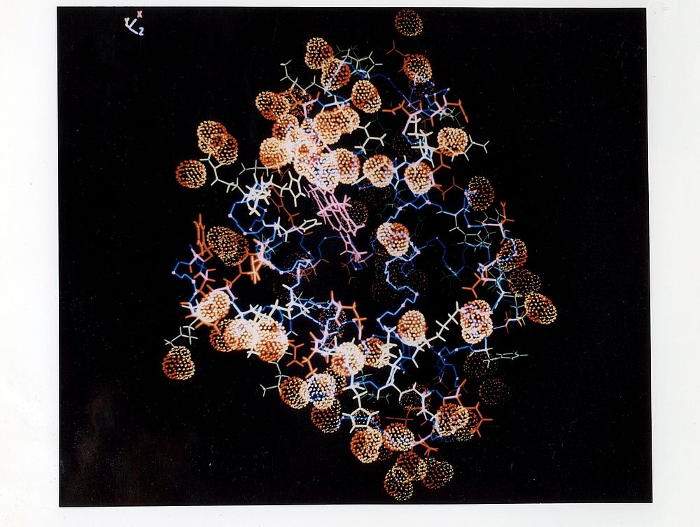
The power of neutron diffraction in detecting hydrogen atoms, and thus determining the positions of hydrating water molecules, is shown in this image of hydrated carbon monoxide myoglobin.
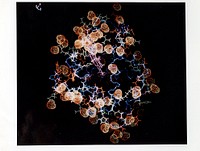
The power of neutron diffraction in detecting hydrogen atoms, and thus determining the positions of hydrating water molecules, is shown in this image of hydrated carbon monoxide myoglobin.
The protein is depicted as a stick model, whereas the water molecules are shown as space-filling stippled structures. Note that the access route (center of picture) to the CO binding site on the pinkish-colored heme group is unobstructed by water molecules. Original public domain image from Flickr
Public DomainFree CCO U.S. Government image for Personal and Business use